Nanopore biosensors for precision medicine
Ultra-sensitive, high-throughput biomolecular sensing technologies are transforming basic research and medical diagnostics. For example, recently developed single cell DNA and RNA sequencing methods have been widely used in numerous studies to date, making a huge impact on basic research in life sciences (1). At the forefront of this trend, emerging single-molecule biosensors take a central stage. Specifically, nanopore biosensors are developed as highly accessible and portable devices for extremely accurate studies of genomic, transcriptomics or proteomics information. These studies pave the way towards the development of high precision clinical sensing, for future applications in drug differentiation and personalized medicine.
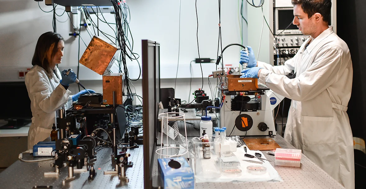
Current studies in our group are focused on isolation and quantification of specific DNA or RNA biomarkers from blood or plasma samples, using nanopore biosensors. The single-molecule level sensitivity of the sensors highly simplifies the sample preparation process required for sensing, and sharply reduces the amount of clinical sample required. Multiplexing of many different biomarkers (such as single-nucleotide variations) is achieved by specific ligation of DNA barcodes, composed of multiple color tags. The readout of the fluorescent tags is performed by our electro-optical nanopore setup.
DNA translocation through nano-scale pores
Nanopores are essentially single-molecule electrophoretic “gels”. An intense electrical field attracts and threads the electrically-charged DNA strand through a pore embedded in an insulating membrane separating two reservoirs filled with ionic aqueous solutions. The nanopore size is only slightly larger than the biopolymer cross section, hence the molecule unfolds and translocates in a single-file manner through it. The electrical field pulling the DNA through the pore also generates an ion current that can be measured as a function of time. During the passage of the DNA through the pore it partly blocks the ionic current, giving rise to a detectable “resistive pulsing” signals.
Electro-optical sensing in Nanopores
Multi-color, single-molecule fluorescence can substantially expand the sensing capabilities of nanopores by complementing or substituting the resistive pulsing signals. Over the past decade our lab developed nanopore biosensors equipped with simultaneous single-photon detectors. This has allowed us to quantify epi-genetic markers such as methylated cytosines in genomic DNA, resolve DNA barcodes for genotyping applications and distinguish among polypeptides, at the single molecule level.


