
Single Molecule Biophysics
and Bio-nano Technology
Welcome to the Meller lab
Department of Biomedical Engineering, Technion
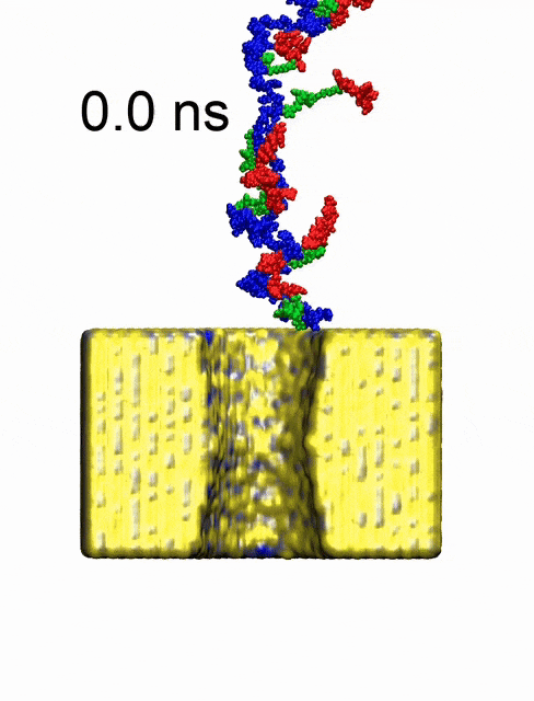
All-atoms molecular dynamics clip showing a single file threading of cysteines conjugated protein through a 4 nm solid-state nanopores under the influence of an electrical field. Movie taken from Soni, N. et al. Nat. Nanotechnol. 2025, 20 (10), 1482–1490.
Movie credit: A. Aksimentiev’s lab, UIUC
Full-length protein classification via cysteine fingerprinting in solid-state nanopores

On-chip separation and imaging of individual proteins
Our lab manages to produce a silicon-based nanofluidic device that allows single-molecule resolution SDS PAGE in sub-micrometer channels. It enables identification, tracking, and analysis of intensity and mobility of single proteins in the sample. This device will permit a wide range of high resolution of protein separations including clinical sample analysis for future medical diagnostics applications.

Stretching and threading ultra-long genomic DNA
A 400 kilobase pair genomic DNA stretched and threaded through a nanoscale pore.
The device is described in a recent paper that appeared in ACS Nano (November, 22 2019): On-Chip Stretching, Sorting, and Electro-Optical Nanopore Sensing of Ultra-Long Human Genomic DNA, Adam Zrehen, Diana Huttner and Amit Meller.
DOI: 10.1021/acsnano.9b07873

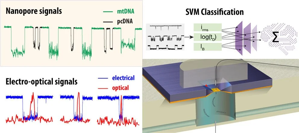
Nanopore biosensors for precision medicine
Nanopores are novel single molecule biosensors. They utilize electrophoretic forces to funnel and thread electrically-charged biomolecules through nano-meter scale pores made in ultra-thin membranes. Our lab has developed and used Nanopore sensors over the past two decades, for a wide range of biomedical applications, including DNA sequencing, RNA secondary-structure determination, DNA interactions with transcription factors, DNA epigenetic modification quantification, and single protein sensing.

Dynamic processes in live cells
Super-resolution microscopy allows quantification of dynamic processes in fixed and live cells. Our lab studies the protein translation initiation machinery — the rate limiting step for ribosome assembly and protein synthesis in all eukaryotic cells.
To image these molecular processes in their native environment, the living cell, new types of imaging methods are being developed that allow sub-diffraction limit resolution with high temporal resolution. Specifically, we employ a custom made parallel STED microscope that allows interrogation of cellular process with unprecedented resolutions.
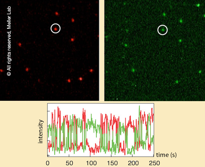
Single molecule spectroscopy
Single molecule FRET (Förster Resonance Energy Transfer) can be used as a spectroscopic ruler to report on inter- and intra-molecular distance changes in real-time. In the past we used smFRET to monitor RNA helices kinetics, distances along dsDNA, and the blinking phenomena of organic dyes. In collaboration with Prof. Oded Lewinson at Technion, we currently study the conformational dynamics of a central bacterial transporter BtuCDF.
News